As already mentioned nervous coordination is brought about through the various elements of the nervous system which are:
(i) Receptors (iv) Motor neurons or nerves
(ii) Sensory neurons or nerves (v) Effectors organs
(iii) Central neurons system (brain and spinal cord), made of associative neurons (inter-neurons)
The stimuli are received by the receptors which convey the messages to the CNS through sensory neurons or nerves. CNS consolidates the information of stimuli, comprehends it and formulates the type of response to be produced. The messages of the type of response are passed via motor neurons or nerves to the particular effectors which produce a specific response.
Nervous coordination is usually involved in producing rapid and short lived responses.
Components of nervous system
1. Neurons (nerve cells)
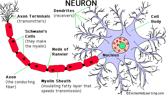
The structural and functional unit of the nervous system in all animals including man is the neuron. This is a highly specialized cell which contains the typical organelles found in most eukaryotic cells. This cell is highly adapted for communication because of its wire like projection; the dendrites which are often further branched and carry impulses towards the central cell body. The cell body is a thicker region of the neuron containing the nucleus and most of the cytoplasm. The axon is the projection, generally very long, that carries impulses away from the cell body. Usually a neuron has a single axon. Fatty substances covering the axon is the myelin sheath along with short regions of exposed axon are called nodes of Ranvier. Many axons and even dendrites combine to form a single nerve.
Neurons are of three types.
a)Sensory neurons: they carry nerve impulses (message from receptors to central nervous system.
b)Motor neurons: they carry nerve impulse (orders) from central nervous system to effectors.
c)Associated neurons: they form central nervous system and are responsible for analyzing the message and issuing orders.
“Examine a photograph/slide of a neuron and compare its structure with Fig 15.1”.

Brain
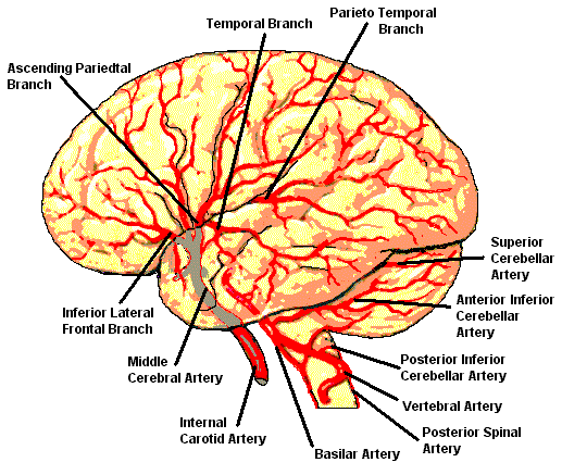
The brain is encased in a bony shell and is further protected by three membranes or meanings. The inner part of these covers the brain and is richly supplied with blood vessels. It brings oxygen and nutrition to underlying brain and protects it. The cerebrospinal fluid (CSF) lies between the inner and middle layers, which, provides cushioning and ions to the brain and spinal cord. The outer most layer is tough and fibrous and provide mechanical support.
The vertebrate brain is divided into three basic regions, the hindbrain, the midbrain and the forebrain (fig 15.2)

The vertebrate brain is chiefly concerned with involuntary, mechanical process. It consists of three primary structures. (1) The medulla oblongata which lies on top of the spinal cord and contains many of the centers that control involuntary process like breathing, blood pressure and heart beat. All communication between brain and spinal cord pass through the medulla. (2) The pons that is present on top of the medulla and contains the longitudinal bundles of myelinated fibres running between brain and spinal cord and controls balance and muscle coordination. (3) The cerebellum lies behind the medulla and controls balance and muscle coordination.
“The human brain is like a super performing a variety of activities. Such as receiving information then interpreting it, passing messages and storing memory”.
The midbrain lies between the hindbrain and forebrain and connects the two. It processes the visual and auditory information from the eyes and ears before sending them to the forebrain.
The forebrain is most advanced in human. Its lower most part which lies above the midbrain is called hypothalamus which control, heart rate, blood pressure, body temperature, hunger, thirst, sex and anger in addition to its hormonal role. Resting above the hypothalamus is the thalamus which provides connections between many parts of brain and between sensory system and cerebrum. It may also control moods and feelings. Sleep may be influenced by thalamus centre along with the midbrain and hindbrain. It is divided into two hemispheres comprising 15 billion nerve cells. Each hemisphere is divided into four major lobes.
(i)At the back are the occipital lobes which receive and analyze visual information’s.
(ii)At the lower sides of the brain are the temporal lobe which are primarily concerned with hearing.
(iii) At the front of the brain are the frontal lobes which regulate fine motor control including movements involved in speech.
Above the front of the brain are the frontal lobes which receive stimuli from the skin and also provide awareness of the body position. Memory track are also supposed to be present in the cerebrum.
Spinal cord
Spinal cord is a thick dorsal neural track extending from the brainstem to the lower back. It is completely enclosed by the vertebrae of the vertebral column just as the brain is enclosed by the bones of the skull. Most of the spinal cord consists of ascending track conducting sensory impulse towards the brain and descending track carrying impulses down ward to motor neurons within the spinal cord. At every level the spinal cord also serves as a reflex center possibly involving a variable numbers of inter neurons. There are 31 pairs of spinal nerves arising from the spinal cord. Spinal cord is concerned with spinal reflex actions and also serves as pathway of information from and to the brain.
